Each study has one job
The American Academy of Orthopaedic Surgeons describes the three common studies by what each resolves:
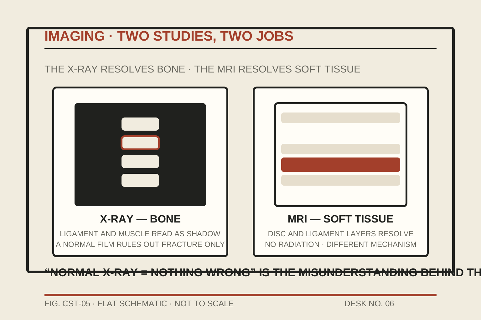
- X-ray (radiography) — bone. Fractures, dislocations, alignment. The standard first study after a crash because it rules out the emergencies; it says nothing about ligament, tendon, or muscle.
- CT — detailed bone, faster and finer than plain films; used where a fracture is suspected but not resolved, or where the joint surface matters.
- MRI — soft tissue. Ligaments, tendons, discs, muscle. No radiation; a different physical mechanism entirely (magnetic resonance, not X-ray).
The normal-X-ray problem
After a crash, the first study is almost always an X-ray — and after a soft-tissue injury, that X-ray is typically normal. That is correct and expected: a normal X-ray rules out fracture, which is its job. It does not rule out injury. The misunderstanding that "normal X-ray = nothing wrong" is one of the three forces behind the underestimation problem.
When MRI enters the picture
MRI is not a routine first step. It is ordered when the clinical picture asks for soft-tissue resolution — persistent pain beyond the expected course, neurological signs on exam, or a suspected disc or ligament injury that the exam cannot settle. The ordering decision is the clinician's, made from the exam and the record; this desk states the structure, not the indication for any individual.
The studies date and describe the injury; they do not feel it for you. The documentation file explains where the films and reports live in the evidence file.